The subtopic in the middle and twelve tasks round it: label four diagrams, complete two tables, put two processes in order and fill 61 gaps. Every standard level statement has a box; the two boxes for higher level only are dashed and badged HL, and drop off for an SL class. Type into the blanks and check them, or print it on A4, Letter or A3 with an answer key.
Paper
Each subtopic on a landscape page, as shuffled and chosen on screen.
A2.2
Cell structure
Cells · Unity and diversity
Hover over a box to see it. Untick it to leave it off this sheet and its printout.
Name
A2.2.2Under the microscope
Number the steps in order.
Focus on the cells at low power with the coarse focus knob.
Lower a cover slip at an angle, so no air bubbles are trapped.
Measure a cell at this magnification with a calibrated eyepiece graticule.
Turn to a higher-power objective lens and use only the fine focus.
Place a thin layer of cells on a slide in a drop of stain.
Magnification = image size ÷ . A drawing 45 mm long of a cell that is really 30 µm long is magnified times. A beside an image shows the real length a line stands for.
A2.2.7Life in one cell
A unicellular organism such as Paramecium carries out every function of life in one cell. It takes in food (), keeps its internal conditions within limits (), carries out all its chemical reactions (), gets rid of the waste those reactions make (), and also moves, grows, responds to stimuli and reproduces.
A2.2.8, A2.2.9Animals, fungi and plants
Plant cells have walls of , fungal cells walls of , and animal cells no wall. Of the three, only plants have chloroplasts and other plastids, and a large, permanent sap vacuole that keeps the cell turgid; any vacuoles in animal cells are small and temporary. Most animal cells have , and some have cilia or flagella, which most fungi and plants lack. Most cells have one nucleus, but aseptate fungal hyphae and skeletal muscle fibres have , while mammal red blood cells and phloem sieve tubes have .
A2.2.12Cells inside cellsHL
Number the steps in order.
Both gain: the host gets energy from the bacterium's aerobic respiration, and the bacterium gets food and shelter.
The bacterium is not digested, and lives on inside the host cell.
Over many generations the bacterium loses its independence and becomes a mitochondrion.
A single-celled eukaryote, which already has a nucleus, engulfs an aerobic bacterium.
Later, some eukaryotes take in a photosynthetic bacterium in the same way: the first chloroplasts.
Mitochondria and chloroplasts still have their own ribosomes and naked, DNA, and new ones arise only by of existing ones, as bacteria do. A theory is strong when it explains a wide range of .
A2.2.5, A2.2.10A bacterium
12345
1
2
3
4
5
Unlike a eukaryote cell, a prokaryote has no , and its DNA is not bound to .
A2.2.10, A2.2.11What the parts do
Structure
In an electron micrograph
Function
Rough ER
flattened sacs dotted with ribosomes
makes for secretion
Smooth ER
tubes and sacs with a smooth surface
makes
Golgi apparatus
a stack of curved, flattened sacs
the rough ER's products and sorts them into vesicles
Secretory vesicle
a small sac near the plasma membrane
releases its contents by
Microvilli
finger-like projections of the plasma membrane
increase the for absorption
A2.2Cell structure
A2.2.13, A2.2.14Many cells, many jobsHL
All the cells of a multicellular organism carry the same genes, yet they into many types. The basis is different patterns of , often triggered by changes in a cell's . Multicellularity has evolved : all plants and animals, and many fungi and algae, are multicellular. It allows a body, with different cells doing different jobs.
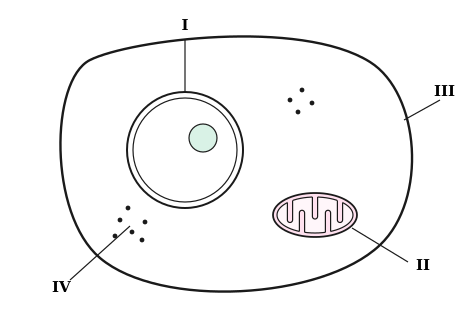
A2.2 examAn animal cell as seen with an electron microscope
1234
1
2
3
4
A2.2.1, A2.2.4What every cell has
Cell theory says that all living organisms are made of one or more cells. So when a new organism is discovered, we can predict that it too is made of cells: reasoning from a theory. Every cell has as its genetic material; a , mostly water, in which its reactions happen; and a made of , which keeps the inside separate from the outside.
A2.2.3Beyond the light microscope
Technique
What it offers
Electron microscopy
a beam of electrons gives far higher
Freeze fracture
frozen cells are split along the middle of their , showing the proteins inside
Cryogenic electron microscopy
fast freezing lets the 3D shapes of protein molecules be seen
Fluorescent stains
dyes bind to chosen structures and glow, each in its own
Immunofluorescence
carry a fluorescent dye to one particular protein
A2.2.6, A2.2.10An animal cell
12345
1
2
3
4
5
Membranes divide the cytoplasm into , the DNA lies within a double membrane with , the ribosomes are the larger type, and a of microtubules and microfilaments supports the cell.
A2.2.8, A2.2.10Identify this cell
12345
1
2
3
4
5
Together, these parts show it is a cell.
Word bank150070S70S ribosome80Sactual sizeantibodiescell wallcellulosecentrioleschitinchloroplastcircularcolourcompartmentscytoplasmcytoskeletondeductivedifferentiatedivisionDNAenvironmentexcretionexocytosisgene expressionGolgi apparatushistoneshomeostasislargerlipidslysosomemanymembranesmetabolismmitochondrionmodifiesnonenucleoidnucleusnutritionobservationsplantplasma membraneporesproteinsrepeatedlyresolutionribosomesrough ERsap vacuolescale barsurface area
Answer key
Every box on the sheet with its gaps filled in, as the subtopic reads in full.
GapsWhat every cell has
Cell theory says that all living organisms are made of one or more cells. So when a new organism is discovered, we can predict that it too is made of cells: deductive reasoning from a theory. Every cell has DNA as its genetic material; a cytoplasm, mostly water, in which its reactions happen; and a plasma membrane made of lipids, which keeps the inside separate from the outside.
OrderUnder the microscope
Place a thin layer of cells on a slide in a drop of stain.
Lower a cover slip at an angle, so no air bubbles are trapped.
Focus on the cells at low power with the coarse focus knob.
Turn to a higher-power objective lens and use only the fine focus.
Measure a cell at this magnification with a calibrated eyepiece graticule.
Magnification = image size ÷ actual size. A drawing 45 mm long of a cell that is really 30 µm long is magnified 1500 times. A scale bar beside an image shows the real length a line stands for.
TableBeyond the light microscope
Technique
What it offers
Electron microscopy
a beam of electrons gives far higher resolution
Freeze fracture
frozen cells are split along the middle of their membranes, showing the proteins inside
Cryogenic electron microscopy
fast freezing lets the 3D shapes of protein molecules be seen
Fluorescent stains
dyes bind to chosen structures and glow, each in its own colour
Immunofluorescence
antibodies carry a fluorescent dye to one particular protein
LabelA bacterium
cell wall
plasma membrane
cytoplasm
nucleoid
70S ribosome
Unlike a eukaryote cell, a prokaryote has no nucleus, and its DNA is not bound to histones.
LabelAn animal cell
nucleus
mitochondrion
rough ER
Golgi apparatus
lysosome
Membranes divide the cytoplasm into compartments, the DNA lies within a double membrane with pores, the ribosomes are the larger 80S type, and a cytoskeleton of microtubules and microfilaments supports the cell.
GapsLife in one cell
A unicellular organism such as Paramecium carries out every function of life in one cell. It takes in food (nutrition), keeps its internal conditions within limits (homeostasis), carries out all its chemical reactions (metabolism), gets rid of the waste those reactions make (excretion), and also moves, grows, responds to stimuli and reproduces.
GapsAnimals, fungi and plants
Plant cells have walls of cellulose, fungal cells walls of chitin, and animal cells no wall. Of the three, only plants have chloroplasts and other plastids, and a large, permanent sap vacuole that keeps the cell turgid; any vacuoles in animal cells are small and temporary. Most animal cells have centrioles, and some have cilia or flagella, which most fungi and plants lack. Most cells have one nucleus, but aseptate fungal hyphae and skeletal muscle fibres have many, while mammal red blood cells and phloem sieve tubes have none.
LabelIdentify this cell
cell wall
chloroplast
sap vacuole
nucleus
mitochondrion
Together, these parts show it is a plant cell.
TableWhat the parts do
Structure
In an electron micrograph
Function
Rough ER
flattened sacs dotted with ribosomes
makes proteins for secretion
Smooth ER
tubes and sacs with a smooth surface
makes lipids
Golgi apparatus
a stack of curved, flattened sacs
modifies the rough ER's products and sorts them into vesicles
Secretory vesicle
a small sac near the plasma membrane
releases its contents by exocytosis
Microvilli
finger-like projections of the plasma membrane
increase the surface area for absorption
OrderCells inside cells HL
A single-celled eukaryote, which already has a nucleus, engulfs an aerobic bacterium.
The bacterium is not digested, and lives on inside the host cell.
Both gain: the host gets energy from the bacterium's aerobic respiration, and the bacterium gets food and shelter.
Over many generations the bacterium loses its independence and becomes a mitochondrion.
Later, some eukaryotes take in a photosynthetic bacterium in the same way: the first chloroplasts.
Mitochondria and chloroplasts still have their own 70S ribosomes and naked, circular DNA, and new ones arise only by division of existing ones, as bacteria do. A theory is strong when it explains a wide range of observations.
GapsMany cells, many jobs HL
All the cells of a multicellular organism carry the same genes, yet they differentiate into many types. The basis is different patterns of gene expression, often triggered by changes in a cell's environment. Multicellularity has evolved repeatedly: all plants and animals, and many fungi and algae, are multicellular. It allows a larger body, with different cells doing different jobs.
Exam diagramAn animal cell as seen with an electron microscope
 Biology by Bradford
 1234
1234