The subtopic in the middle and 14 tasks round it: label seven diagrams, complete two tables, put two processes in order and fill 74 gaps. Every standard level statement has a box; the seven boxes for higher level only are dashed and badged HL, and drop off for an SL class. Type into the blanks and check them, or print it on A4, Letter or A3 with an answer key.
Paper
Each subtopic on a landscape page, as shuffled and chosen on screen.
B3.2
Transport
1 of 2Organisms · Form and function
Hover over a box to see it. Untick it to leave it off this sheet and its printout.
Name
B3.2.6Blocked coronary arteries
Fatty builds up in the wall of a coronary artery and narrows it. A blood forming there can block it completely, so the heart muscle beyond is starved of and dies: a heart attack. Country data like these give r = 0.8, a strong correlation, but a correlation alone cannot prove .
B3.2.9Stem in section
12345
1
2
3
4
5
The vascular bundles lie in a near the outside of the stem. In each, the outer tissue carries and the inner one carries and mineral ions.
B3.2.3, B3.2.4, B3.2.5Artery and vein
Artery
Vein
Blood pressure
low
Wall
thick layers of muscle and tissue
thin and flexible
Keeping blood moving
the stretched wall between beats, keeping the pressure up
stop backflow; nearby muscles squeeze the vein
Each surge that stretches an artery is a pulse. Press two fingertips on the artery at the wrist or the artery in the neck, count for 15 s and multiply by to give beats per minute.
B3.2Transport
B3.2.7Up the xylem
Number the steps in order.
Water evaporates from the cell walls of leaf cells and diffuses out through the stomata.
The tension pulls water up the xylem of the stem as an unbroken column.
This puts the water in the xylem under tension: a negative pressure potential.
Water enters the root from the soil to replace what has moved up.
Water is drawn out of the xylem and through the cell walls by capillary action to replace it.
Water molecules hold on to one another by , so the column does not break.
B3.2.1, B3.2.2Vessels in section
1234
1
2
3
4
The smallest vessels branch into dense beds of narrow tubes with a huge . Their wall is thick, and some have pores, , where exchange must be very fast.
B3.2.8Built to carry water
Xylem vessels are dead tubes with no cell contents and no , so water flows through unimpeded. Their walls are thickened with , so they do not collapse under , and water moves in and out sideways through .
B3.2.10Root in section
1234
1
2
3
4
In a root the vascular tissue sits in the , not in a ring of bundles.
Word bank4arterycapillarycarotidcausationcentreclotcohesioncortexelasticend wallsepidermisfenestrationshighligninlumenone celloxygenphloempithpitsplaquepositiveradialrecoilsringsugarssurface areatensionvalvesveinwaterxylem
B3.2
Transport
2 of 2Organisms · Form and functionName
B3.2.11, B3.2.12, B3.2.13Fluid round the cellsHL
At the arteriole end of a capillary, high blood pressure forces plasma out through the wall: . This tissue fluid has the small solutes of plasma but almost no , which are too large to leave. Cells take O₂ and glucose from it and pass CO₂ and wastes into it. At the end the pressure is lower, so most of it drains back in. The excess enters ducts, whose thin walls have gaps and whose keep it flowing back towards the blood.
B3.2.17, B3.2.18Moving sapHL
12345
1
2
3
4
5
Sugars are loaded into the phloem at a , such as a leaf, and unloaded at a , such as a root or fruit. When transpiration is too slow, roots pump mineral ions into the xylem by ; water follows by osmosis, building a positive .
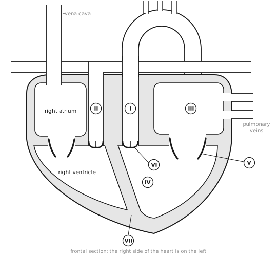
B3.2 examA mammalian heart in the frontal plane (2)HL
123
1
2
3
B3.2.14One circuit or twoHL
Bony fish
Mammal
Route of the blood
heart → → body → heart
heart → → heart → body → heart
Times through the heart in one circuit
once
Pressure of blood reaching the body
high
So a fish has a single circulation and a mammal a one.
B3.2Transport
B3.2.15The heartHL
12345
1
2
3
4
5
The keeps oxygenated and deoxygenated blood apart. The walls are , fed by the , and each beat starts at the heart's own .
B3.2.16One heartbeatHL
Number the steps in order.
Ventricular pressure falls below the atrium's, so the atrioventricular valve opens and the ventricle fills.
The ventricle contracts; its pressure rises above the atrium's, so the atrioventricular valve shuts.
The sinoatrial node fires and the left atrium contracts, topping up the ventricle through the open atrioventricular valve.
The ventricle relaxes; its pressure falls below the aorta's, so the semilunar valve shuts.
Ventricular pressure rises above the aorta's, so the semilunar valve opens and blood is pumped out.
In a blood pressure of 120/80 mmHg, 120 is the pressure, as the ventricles contract, and 80 the pressure, as they relax.
Every box on the sheet with its gaps filled in, as the subtopic reads in full.
LabelVessels in section
artery
vein
lumen
capillary
The smallest vessels branch into dense beds of narrow tubes with a huge surface area. Their wall is one cell thick, and some have pores, fenestrations, where exchange must be very fast.
TableArtery and vein
Artery
Vein
Blood pressure
high
low
Wall
thick layers of muscle and elastic tissue
thin and flexible
Keeping blood moving
the stretched wall recoils between beats, keeping the pressure up
valves stop backflow; nearby muscles squeeze the vein
Each surge that stretches an artery is a pulse. Press two fingertips on the radial artery at the wrist or the carotid artery in the neck, count for 15 s and multiply by 4 to give beats per minute.
GapsBlocked coronary arteries
Fatty plaque builds up in the wall of a coronary artery and narrows it. A blood clot forming there can block it completely, so the heart muscle beyond is starved of oxygen and dies: a heart attack. Country data like these give r = 0.8, a strong positive correlation, but a correlation alone cannot prove causation.
OrderUp the xylem
Water evaporates from the cell walls of leaf cells and diffuses out through the stomata.
Water is drawn out of the xylem and through the cell walls by capillary action to replace it.
This puts the water in the xylem under tension: a negative pressure potential.
The tension pulls water up the xylem of the stem as an unbroken column.
Water enters the root from the soil to replace what has moved up.
Water molecules hold on to one another by cohesion, so the column does not break.
GapsBuilt to carry water
Xylem vessels are dead tubes with no cell contents and no end walls, so water flows through unimpeded. Their walls are thickened with lignin, so they do not collapse under tension, and water moves in and out sideways through pits.
LabelStem in section
epidermis
cortex
phloem
xylem
pith
The vascular bundles lie in a ring near the outside of the stem. In each, the outer tissue carries sugars and the inner one carries water and mineral ions.
LabelRoot in section
epidermis
cortex
xylem
phloem
In a root the vascular tissue sits in the centre, not in a ring of bundles.
GapsFluid round the cells HL
At the arteriole end of a capillary, high blood pressure forces plasma out through the wall: pressure filtration. This tissue fluid has the small solutes of plasma but almost no proteins, which are too large to leave. Cells take O₂ and glucose from it and pass CO₂ and wastes into it. At the venule end the pressure is lower, so most of it drains back in. The excess enters lymph ducts, whose thin walls have gaps and whose valves keep it flowing back towards the blood.
TableOne circuit or two HL
Bony fish
Mammal
Route of the blood
heart → gills → body → heart
heart → lungs → heart → body → heart
Times through the heart in one circuit
once
twice
Pressure of blood reaching the body
low
high
So a fish has a single circulation and a mammal a double one.
LabelThe heart HL
right atrium
left ventricle
atrioventricular valve
semilunar valve
aorta
The septum keeps oxygenated and deoxygenated blood apart. The walls are cardiac muscle, fed by the coronary arteries, and each beat starts at the heart's own pacemaker.
OrderOne heartbeat HL
The sinoatrial node fires and the left atrium contracts, topping up the ventricle through the open atrioventricular valve.
The ventricle contracts; its pressure rises above the atrium's, so the atrioventricular valve shuts.
Ventricular pressure rises above the aorta's, so the semilunar valve opens and blood is pumped out.
The ventricle relaxes; its pressure falls below the aorta's, so the semilunar valve shuts.
Ventricular pressure falls below the atrium's, so the atrioventricular valve opens and the ventricle fills.
In a blood pressure of 120/80 mmHg, 120 is the systolic pressure, as the ventricles contract, and 80 the diastolic pressure, as they relax.
LabelMoving sap HL
sieve tube element
sieve plate
companion cell
mitochondrion
plasmodesmata
Sugars are loaded into the phloem at a source, such as a leaf, and unloaded at a sink, such as a root or fruit. When transpiration is too slow, roots pump mineral ions into the xylem by active transport; water follows by osmosis, building a positive root pressure.
Exam diagramA mammalian heart in the frontal plane HL
aorta
pulmonary artery
left atrium
left ventricle
Exam diagramA mammalian heart in the frontal plane (2) HL
 Biology by Bradford
 123
123