D2.1.8Name the phase
1234
- 1
- 2
- 3
- 4
 Biology by Bradford
support
Biology by Bradford
support
The subtopic in the middle and twelve tasks round it: label five diagrams, complete two tables, put a process in order and fill 63 gaps. Every standard level statement has a box; the four boxes for higher level only are dashed and badged HL, and drop off for an SL class. Type into the blanks and check them, or print it on A4, Letter or A3 with an answer key.
Early in meiosis I, homologous chromosomes pair up as a . chromatids cross at a and swap matching sections, giving new combinations of alleles. Then each pair lines up facing either pole by chance, , so a cell with n pairs can give combinations of chromosomes in its gametes: over 8 million for the 23 pairs in a human.
Cells proliferate by this cycle: for growth, in plant and early embryos, and to replace cells lost from the and heal its wounds. Interphase is a busy time: the cell makes proteins and other components, and its grow and divide, as do chloroplasts in a plant.
A mutation can turn a gene that stimulates division into an , which keeps division switched on, or knock out a gene that normally holds it back. Cells then divide out of control, forming a tumour. A tumour is a cancer: it invades neighbouring tissue, and cells breaking away from this primary tumour can form tumours elsewhere, . A benign tumour grows more slowly and stays where it is.
| Mitosis | Meiosis | |
|---|---|---|
| Divisions of the nucleus | one | |
| From one diploid nucleus | two diploid nuclei | four nuclei |
| Chromosome number | halved | |
| Daughter nuclei are genetically | ||
| Used for | growth, repair, asexual reproduction | , in a sexual life cycle |
The nucleus divides before the cell does, so that no daughter cell is left , with no nucleus at all.
Before mitosis or meiosis every DNA molecule is , and the two copies stay held together until . The DNA winds round and then coils on itself again and again, , making a short, thick chromosome that can be moved without tangling. Motor proteins do the moving.
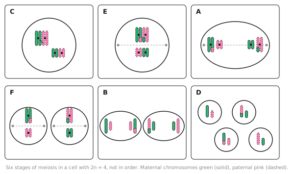
Meiosis has two divisions. In the first, the two chromosomes of each pair go to opposite poles; in the second, the sister separate. If a pair fails to separate, , a gamete ends up with an extra chromosome. A child from such a gamete with three copies of chromosome has .
Proteins called rise and fall in concentration through the cell cycle. A cell can pass each only when a particular one reaches a level; that one then again.
Cytokinesis usually splits the cytoplasm , but not in human , where one cell keeps nearly all of it, nor in yeast, which reproduces by . Every daughter cell needs at least one , as these can only be made by the division of existing ones.
Number the steps in order.
Each new nucleus gets one chromatid of every chromosome, so the two are genetically .
| Sample | Cells in mitosis | Cells counted | Mitotic index |
|---|---|---|---|
| Normal skin | 6 | 200 | |
| Benign tumour | 200 | 0.10 | |
| Malignant tumour | 40 | 160 |
Mitotic index = number of cells in mitosis ÷ . Cells in mitosis are picked out by their visible, condensed .
 1234
1234Cytokinesis usually splits the cytoplasm equally, but not in human oogenesis, where one cell keeps nearly all of it, nor in yeast, which reproduces by budding. Every daughter cell needs at least one mitochondrion, as these can only be made by the division of existing ones.
| Mitosis | Meiosis | |
|---|---|---|
| Divisions of the nucleus | one | two |
| From one diploid nucleus | two diploid nuclei | four haploid nuclei |
| Chromosome number | maintained | halved |
| Daughter nuclei are genetically | identical | different |
| Used for | growth, repair, asexual reproduction | gametes, in a sexual life cycle |
The nucleus divides before the cell does, so that no daughter cell is left anucleate, with no nucleus at all.
Before mitosis or meiosis every DNA molecule is replicated, and the two copies stay held together until anaphase. The DNA winds round histones and then coils on itself again and again, supercoiling, making a short, thick chromosome that can be moved without tangling. Motor proteins do the moving.
Each new nucleus gets one chromatid of every chromosome, so the two are genetically identical.
Meiosis has two divisions. In the first, the two chromosomes of each homologous pair go to opposite poles; in the second, the sister chromatids separate. If a pair fails to separate, non-disjunction, a gamete ends up with an extra chromosome. A child from such a gamete with three copies of chromosome 21 has Down syndrome.
Early in meiosis I, homologous chromosomes pair up as a bivalent. Non-sister chromatids cross at a chiasma and swap matching sections, giving new combinations of alleles. Then each pair lines up facing either pole by chance, random orientation, so a cell with n pairs can give 2ⁿ combinations of chromosomes in its gametes: over 8 million for the 23 pairs in a human.
Cells proliferate by this cycle: for growth, in plant meristems and early embryos, and to replace cells lost from the skin and heal its wounds. Interphase is a busy time: the cell makes proteins and other components, and its mitochondria grow and divide, as do chloroplasts in a plant.
Proteins called cyclins rise and fall in concentration through the cell cycle. A cell can pass each checkpoint only when a particular one reaches a threshold level; that one then falls again.
A mutation can turn a gene that stimulates division into an oncogene, which keeps division switched on, or knock out a tumour suppressor gene that normally holds it back. Cells then divide out of control, forming a tumour. A malignant tumour is a cancer: it invades neighbouring tissue, and cells breaking away from this primary tumour can form secondary tumours elsewhere, metastasis. A benign tumour grows more slowly and stays where it is.
| Sample | Cells in mitosis | Cells counted | Mitotic index |
|---|---|---|---|
| Normal skin | 6 | 200 | 0.03 |
| Benign tumour | 20 | 200 | 0.10 |
| Malignant tumour | 40 | 160 | 0.25 |
Mitotic index = number of cells in mitosis ÷ total number of cells. Cells in mitosis are picked out by their visible, condensed chromosomes.