The subtopic in the middle and eleven tasks round it: label four diagrams, complete three tables, put two processes in order and fill 48 gaps. Every standard level statement has a box; the five boxes for higher level only are dashed and badged HL, and drop off for an SL class. Type into the blanks and check them, or print it on A4, Letter or A3 with an answer key.
Paper
Each subtopic on a landscape page, as shuffled and chosen on screen.
D3.3
Homeostasis
Organisms · Continuity and change
Hover over a box to see it. Untick it to leave it off this sheet and its printout.
Name
D3.3.3After a sugary meal
Number the steps in order.
Insulin is carried in the blood to target cells all over the body.
Liver and muscle cells take up glucose and store it as glycogen.
Beta cells in the pancreas detect the rise and secrete insulin.
Glucose absorbed from the gut raises blood glucose concentration.
Blood glucose falls back to the set point, and insulin secretion falls.
When blood glucose falls too low, secrete , which makes liver cells break glycogen down to glucose.
D3.3.7, D3.3.9Two jobs of the kidneyHL
is the removal of waste products of metabolism, such as urea. is control of the osmotic concentration of body fluids, measured in osmol L⁻¹. In the loop of Henle, the ascending limb pumps out by , keeping the osmotic concentration of the medulla high so water can be reabsorbed from the collecting ducts.
D3.3.8, D3.3.9, D3.3.10Inside a nephronHL
12345
1
2
3
4
5
Blood pressure forces water and small solutes out of the plasma: . Then all the glucose and amino acids, and most of the water, are , leaving and other unwanted solutes to be excreted in urine.
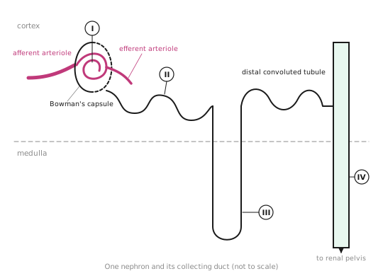
D3.3 examA nephron and its collecting ductHL
1234
1
2
3
4
D3.3.4Two types of diabetes
Type 1
Type 2
Cause
the immune system destroys the
target cells become to insulin
Usual onset
childhood
adulthood
Risk factors
inherited genes
, inactivity, family history
Treatment or prevention
insulin injections, with blood glucose checks
a healthy diet and , then drugs if needed
D3.3.5Temperature control loop
1234
1
2
3
4
The gland in the neck secretes , which raises the so body cells release more heat. Muscle and adipose tissue are , carrying out the response.
D3.3Homeostasis
D3.3.6Skin in heat and cold
123
1
2
3
Birds and mammals regulate their temperature by physiological changes and by , such as seeking shade or huddling together.
D3.3.1, D3.3.2Keeping things steady
Homeostasis keeps the within preset limits, whatever happens outside. In humans, body temperature, blood pH, blood glucose concentration and blood are all held near a . feedback brings a variable back whether it rises above or falls below; positive feedback would push it further away.
D3.3.6Too hot, too cold
Effector
Response when too cold
Skin arterioles
: less blood flows near the surface, so less heat is lost
Skeletal muscle
: rapid contractions release heat from respiration
Brown adipose tissue
: energy from respiration is released as heat instead of being used to make ATP
Hair erector muscles
contract, so hairs stand up and trap a layer of insulating
When too hot, skin arterioles widen, , so more heat is lost from the blood at the surface, and sweat takes heat from the skin as it .
D3.3.10Short of waterHL
Number the steps in order.
More water is reabsorbed by osmosis, leaving a small volume of concentrated urine.
The pituitary gland secretes more ADH into the blood.
Vesicles carrying aquaporins fuse with the cell membrane facing the filtrate.
ADH binds to receptors on cells of the collecting duct.
Osmoreceptors in the hypothalamus detect a rise in blood osmotic concentration.
With plenty of water, ADH secretion falls, the aquaporins are taken back into the cells, and a large volume of urine forms.
D3.3.11Where the blood goesHL
Organ
Blood flow in vigorous exercise, compared with rest
Reason
Skeletal muscles
arterioles widen to bring oxygen and glucose for respiration
Gut
arterioles narrow; digestion can wait
Kidneys
arterioles narrow, sending blood to the muscles
Brain
it must never be short of oxygen
Word bankactive transportairalpha cellsarterioleascending limb of the loop of Henlebehaviourbeta cellsBowman's capsulecollecting ductdecreasesdiluteeffectorsevaporatesExcretionexerciseglomerulusglucagonhair erector musclehypothalamusincreasesinternal environmentloop of Henlemetabolic rateNegativeobesityOsmoregulationosmotic concentrationpituitary glandproximal convoluted tubulereabsorbedresistantset pointshiveringsodium ionsstays the samesweat glandthermoreceptorthyroid glandthyroxinultrafiltrationuncoupled respirationureavasoconstrictionvasodilation
Answer key
Every box on the sheet with its gaps filled in, as the subtopic reads in full.
GapsKeeping things steady
Homeostasis keeps the internal environment within preset limits, whatever happens outside. In humans, body temperature, blood pH, blood glucose concentration and blood osmotic concentration are all held near a set point. Negative feedback brings a variable back whether it rises above or falls below; positive feedback would push it further away.
OrderAfter a sugary meal
Glucose absorbed from the gut raises blood glucose concentration.
Beta cells in the pancreas detect the rise and secrete insulin.
Insulin is carried in the blood to target cells all over the body.
Liver and muscle cells take up glucose and store it as glycogen.
Blood glucose falls back to the set point, and insulin secretion falls.
When blood glucose falls too low, alpha cells secrete glucagon, which makes liver cells break glycogen down to glucose.
TableTwo types of diabetes
Type 1
Type 2
Cause
the immune system destroys the beta cells
target cells become resistant to insulin
Usual onset
childhood
adulthood
Risk factors
inherited genes
obesity, inactivity, family history
Treatment or prevention
insulin injections, with blood glucose checks
a healthy diet and exercise, then drugs if needed
LabelTemperature control loop
thermoreceptor
hypothalamus
pituitary gland
thyroid gland
The gland in the neck secretes thyroxin, which raises the metabolic rate so body cells release more heat. Muscle and adipose tissue are effectors, carrying out the response.
LabelSkin in heat and cold
arteriole
hair erector muscle
sweat gland
Birds and mammals regulate their temperature by physiological changes and by behaviour, such as seeking shade or huddling together.
TableToo hot, too cold
Effector
Response when too cold
Skin arterioles
vasoconstriction: less blood flows near the surface, so less heat is lost
Skeletal muscle
shivering: rapid contractions release heat from respiration
Brown adipose tissue
uncoupled respiration: energy from respiration is released as heat instead of being used to make ATP
Hair erector muscles
contract, so hairs stand up and trap a layer of insulating air
When too hot, skin arterioles widen, vasodilation, so more heat is lost from the blood at the surface, and sweat takes heat from the skin as it evaporates.
GapsTwo jobs of the kidney HL
Excretion is the removal of waste products of metabolism, such as urea. Osmoregulation is control of the osmotic concentration of body fluids, measured in osmol L⁻¹. In the loop of Henle, the ascending limb pumps sodium ions out by active transport, keeping the osmotic concentration of the medulla high so water can be reabsorbed from the collecting ducts.
LabelInside a nephron HL
glomerulus
Bowman's capsule
proximal convoluted tubule
loop of Henle
collecting duct
Blood pressure forces water and small solutes out of the plasma: ultrafiltration. Then all the glucose and amino acids, and most of the water, are reabsorbed, leaving urea and other unwanted solutes to be excreted in urine.
OrderShort of water HL
Osmoreceptors in the hypothalamus detect a rise in blood osmotic concentration.
The pituitary gland secretes more ADH into the blood.
ADH binds to receptors on cells of the collecting duct.
Vesicles carrying aquaporins fuse with the cell membrane facing the filtrate.
More water is reabsorbed by osmosis, leaving a small volume of concentrated urine.
With plenty of water, ADH secretion falls, the aquaporins are taken back into the cells, and a large volume of dilute urine forms.
TableWhere the blood goes HL
Organ
Blood flow in vigorous exercise, compared with rest
Reason
Skeletal muscles
increases
arterioles widen to bring oxygen and glucose for respiration
 Biology by Bradford
 1234
1234