B2.1 Membranes and membrane transport: revision map
The subtopic in the middle and eleven tasks round it: label five diagrams, complete a table, put a process in order and fill 57 gaps. Every standard level statement has a box; the four boxes for higher level only are dashed and badged HL, and drop off for an SL class. Type into the blanks and check them, or print it on A4, Letter or A3 with an answer key.
Paper
Each subtopic on a landscape page, as shuffled and chosen on screen.
B2.1
Membranes and membrane transport
Cells · Form and function
Hover over a box to see it. Untick it to leave it off this sheet and its printout.
Name
B2.1.9Carbohydrate tags
Short carbohydrate chains are attached to some membrane proteins, making , and to some lipids, making . The chains are only ever on the side of the plasma membrane. Their shapes act as markers in cell , so the immune system can tell self from non-self, and they help neighbouring cells stick together in cell .
B2.1.3, B2.1.6, B2.1.7, B2.1.8Ways across a membrane
Route across
Concentration gradient
Simple diffusion
between the phospholipids
Facilitated diffusion
through
Active transport
through proteins that use energy
Channels and pumps each pass only particular particles, so they make a membrane selective. Simple diffusion is not: which particles cross depends only on their and on whether they are hydrophilic or hydrophobic. O₂ and CO₂ cross this way, being small and .
B2.1.11, B2.1.12Fluid at any temperatureHL
Kinked fatty acid tails pack loosely and have lower melting points, keeping a membrane fluid and flexible at the temperatures a cell meets. Straight tails pack tightly and make a membrane stronger at higher temperatures; fish of polar seas have more of the kinked kind in their membranes. In animal cells, sits among the phospholipids and adjusts fluidity: it the membrane when warm and stops it becoming when cold.
B2.1.14, B2.1.15An exchange transporterHL
Number the steps in order.
Three Na⁺ ions from the cytoplasm bind to the pump.
The pump changes shape, opens to the outside and releases the Na⁺.
Two K⁺ ions from outside bind to the pump.
The phosphate group leaves and the pump returns to its first shape.
The K⁺ ions are released into the cytoplasm.
ATP transfers a phosphate group to the pump.
Each turn moves more positive charge out than in, helping to make the inside of a neuron compared with the outside: a membrane potential. Na⁺ and K⁺ channels in neurons open when that potential changes, so they are -gated; nicotinic receptors open when binds, so they are neurotransmitter-gated.
B2.1.13Budding and fusingHL
123
1
2
3
Both rely on the membrane being fluid, so that it can pinch apart and join up again without leaking. A takes in bacteria the way shown on the left; a pancreas cell releases insulin the way shown on the right.
B2.1 examA section of a plasma membrane
1234
1
2
3
4
B2.1Membranes and membrane transport
B2.1.15, B2.1.16, B2.1.17Absorbing glucoseHL
123
1
2
3
Na⁺ is kept at a concentration inside the cell, so it diffuses in from the gut, and glucose is carried in with it its own concentration gradient. ATP is not used at this step, so this is active transport. Cells of the nephron use the same method to glucose from the filtrate.
B2.1.1, B2.1.2Lipids meet water
Phospholipids are : each has a hydrophilic phosphate head and two hydrophobic tails. In water they form a continuous , heads facing the water on each side and tails meeting in the core. That core lets very few large molecules, or polar molecules through, so the membrane is an effective between two watery solutions.
B2.1.4, B2.1.10The fluid mosaic model
12345
1
2
3
4
5
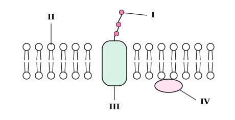
Fluid: the lipids and most proteins drift within their own layer. Mosaic: proteins of many kinds lie scattered among the lipids, some carrying particles across, some acting as for signals and some as .
B2.1.5Water on the move
Water molecules move at and cross the membrane in both directions, many of them through protein pores called . The membrane is to the solutes, so they cannot follow, and there is a net movement of water towards the side with the solute concentration: osmosis.
B2.1.6, B2.1.7Through the proteins
123
1
2
3
The left-hand protein has a pore that fits only one kind of ion, and ions diffuse through only while its gate is , so the membrane is permeable. The right-hand protein uses energy from the small molecule beside it to carry particles their concentration gradient.
Every box on the sheet with its gaps filled in, as the subtopic reads in full.
GapsLipids meet water
Phospholipids are amphipathic: each has a hydrophilic phosphate head and two hydrophobic hydrocarbon tails. In water they form a continuous bilayer, heads facing the water on each side and tails meeting in the core. That core lets very few large molecules, ions or polar molecules through, so the membrane is an effective barrier between two watery solutions.
LabelThe fluid mosaic model
phospholipid
cholesterol
integral protein
glycoprotein
peripheral protein
Fluid: the lipids and most proteins drift sideways within their own layer. Mosaic: proteins of many kinds lie scattered among the lipids, some carrying particles across, some acting as receptors for signals and some as enzymes.
GapsCarbohydrate tags
Short carbohydrate chains are attached to some membrane proteins, making glycoproteins, and to some lipids, making glycolipids. The chains are only ever on the extracellular side of the plasma membrane. Their shapes act as markers in cell recognition, so the immune system can tell self from non-self, and they help neighbouring cells stick together in cell adhesion.
GapsWater on the move
Water molecules move at random and cross the membrane in both directions, many of them through protein pores called aquaporins. The membrane is impermeable to the solutes, so they cannot follow, and there is a net movement of water towards the side with the higher solute concentration: osmosis.
LabelThrough the proteins
channel protein
pump protein
ATP
The left-hand protein has a pore that fits only one kind of ion, and ions diffuse through only while its gate is open, so the membrane is selectively permeable. The right-hand protein uses energy from the small molecule beside it to carry particles against their concentration gradient.
TableWays across a membrane
Route across
Concentration gradient
Simple diffusion
between the phospholipids
down it
Facilitated diffusion
through channel proteins
down it
Active transport
through proteins that use energy
against it
Channels and pumps each pass only particular particles, so they make a membrane selective. Simple diffusion is not: which particles cross depends only on their size and on whether they are hydrophilic or hydrophobic. O₂ and CO₂ cross this way, being small and non-polar.
GapsFluid at any temperature HL
Kinked unsaturated fatty acid tails pack loosely and have lower melting points, keeping a membrane fluid and flexible at the temperatures a cell meets. Straight saturated tails pack tightly and make a membrane stronger at higher temperatures; fish of polar seas have more of the kinked kind in their membranes. In animal cells, cholesterol sits among the phospholipids and adjusts fluidity: it stabilises the membrane when warm and stops it becoming stiff when cold.
LabelBudding and fusing HL
endocytosis
vesicle
exocytosis
Both rely on the membrane being fluid, so that it can pinch apart and join up again without leaking. A white blood cell takes in bacteria the way shown on the left; a pancreas cell releases insulin the way shown on the right.
OrderAn exchange transporter HL
Three Na⁺ ions from the cytoplasm bind to the pump.
ATP transfers a phosphate group to the pump.
The pump changes shape, opens to the outside and releases the Na⁺.
Two K⁺ ions from outside bind to the pump.
The phosphate group leaves and the pump returns to its first shape.
The K⁺ ions are released into the cytoplasm.
Each turn moves more positive charge out than in, helping to make the inside of a neuron negative compared with the outside: a membrane potential. Na⁺ and K⁺ channels in neurons open when that potential changes, so they are voltage-gated; nicotinic receptors open when acetylcholine binds, so they are neurotransmitter-gated.
LabelAbsorbing glucose HL
sodium–glucose cotransporter
sodium–potassium pump
cell-adhesion molecules
Na⁺ is kept at a low concentration inside the cell, so it diffuses in from the gut, and glucose is carried in with it against its own concentration gradient. ATP is not used at this step, so this is indirect active transport. Cells of the nephron use the same method to reabsorb glucose from the filtrate.
 Biology by Bradford
 1234
1234