The subtopic in the middle and nine tasks round it: label three diagrams, complete a table, put a process in order and fill 43 gaps. The whole subtopic is higher level. Type into the blanks and check them, or print it on A4, Letter or A3 with an answer key.
Paper
Each subtopic on a landscape page, as shuffled and chosen on screen.
C2.1
Chemical signalling
Cells · Interaction and interdependence
Hover over a box to see it. Untick it to leave it off this sheet and its printout.
Name
C2.1.1, C2.1.7, C2.1.8Binding sitesHL
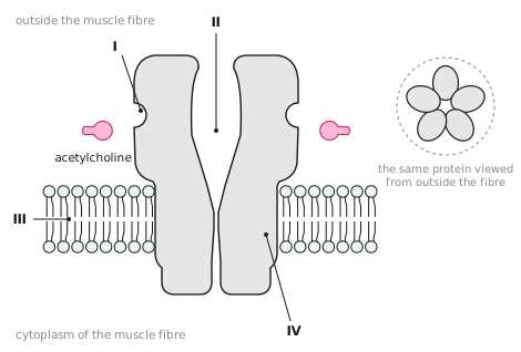
A receptor is a protein with a binding site that fits one particular signalling chemical, its . Binding changes the receptor's , which sets off a pathway: a sequence of responses inside the cell. The acetylcholine receptor is also an ion channel: when it opens, positively charged ions such as diffuse into the cell and change the .
C2.1.6, C2.1.12Inside or outside?HL
1234
1
2
3
4
Where a receptor passes through the membrane, its surface has amino acids. A ligand cannot cross the membrane, so it binds outside. Steroid hormones such as oestradiol, progesterone and testosterone pass straight through and bind inside; the activated receptor binds to specific DNA sequences and promotes of the genes there.
C2.1 examA section through an acetylcholine receptor in the plasmaHL
1234
1
2
3
4
C2.1.3, C2.1.5Four kinds of signalHL
Signal
Released by
Acts on
Hormone
glands
distant cells, carried in the
Neurotransmitter
neurons
the next cell, reached by
Cytokine
many cells, such as immune cells
cells
Calcium ions
channels in membranes
proteins the same cell
C2.1.4Many molecules, many messagesHL
Hormones belong to different chemical groups: epinephrine is an , insulin a , and oestradiol a . Neurotransmitters are just as varied: glutamate is an , the endorphins are , and nitric oxide is a gas. So many different molecules, each with its own receptors, let many separate messages travel at once without being confused.
C2.1Chemical signalling
C2.1.11After a mealHL
1234
1
2
3
4
Binding switches on the receptor's activity inside the cell. A sequence of reactions follows that ends with more carriers in the plasma membrane, so the cell takes in faster.
C2.1.2Bacteria that countHL
Each Vibrio fischeri cell releases a little of a signalling chemical. Where cells are few it diffuses away, but where the population is high, as in the light organ of the bobtail squid, its rises past a threshold. It then binds to in the bacteria, which switch on the genes for all at once. This is .
C2.1.9, C2.1.10Epinephrine's relayHL
Number the steps in order.
cAMP activates protein kinase A, which starts a chain of reactions that breaks glycogen down to glucose in a liver cell.
Adenylyl cyclase converts ATP into cyclic AMP (cAMP).
Epinephrine (adrenaline) binds to a receptor on the outer face of the plasma membrane.
The receptor changes shape and activates a G protein on the inner face, which swaps GDP for GTP.
The G protein switches on adenylyl cyclase, an enzyme in the membrane.
cAMP is the . One epinephrine molecule leads to a great many cAMP molecules, so the signal is . Humans have hundreds of different receptors that work through G proteins.
C2.1.13, C2.1.14Feedback loopsHL
Oestradiol binds to receptors in cells of the that secrete GnRH. Before ovulation, rising oestradiol makes them secrete more GnRH, which leads through the pituitary to still more oestradiol: the response strengthens the signal, so this is feedback. In feedback the response weakens the signal and a level stays steady: insulin lowers blood glucose, so less insulin is then secreted. Progesterone binds to receptors in cells of the , keeping it thick and ready for an embryo to implant.
Word bankamineamino acidamplifiedbinding site for acetylcholinebioluminescencebloodconcentrationdensitydiffusionendocrineendometriumglucoseglucose transporterhydrophilichydrophobichypothalamusinsulinintracellular receptorion channelligandmembrane potentialnearbynegativenucleuspeptidesphosphate groupphospholipid bilayerplasma membranepositiveproteinquorum sensingreceptorssecond messengershapesignal transductionsodiumsteroidtranscriptiontransmembrane receptortransmembrane regiontyrosine kinasevesiclewithin
Answer key
Every box on the sheet with its gaps filled in, as the subtopic reads in full.
GapsBinding sites HL
A receptor is a protein with a binding site that fits one particular signalling chemical, its ligand. Binding changes the receptor's shape, which sets off a signal transduction pathway: a sequence of responses inside the cell. The acetylcholine receptor is also an ion channel: when it opens, positively charged ions such as sodium diffuse into the cell and change the membrane potential.
GapsBacteria that count HL
Each Vibrio fischeri cell releases a little of a signalling chemical. Where cells are few it diffuses away, but where the population density is high, as in the light organ of the bobtail squid, its concentration rises past a threshold. It then binds to receptors in the bacteria, which switch on the genes for bioluminescence all at once. This is quorum sensing.
TableFour kinds of signal HL
Signal
Released by
Acts on
Hormone
endocrine glands
distant cells, carried in the blood
Neurotransmitter
neurons
the next cell, reached by diffusion
Cytokine
many cells, such as immune cells
nearby cells
Calcium ions
channels in membranes
proteins within the same cell
GapsMany molecules, many messages HL
Hormones belong to different chemical groups: epinephrine is an amine, insulin a protein, and oestradiol a steroid. Neurotransmitters are just as varied: glutamate is an amino acid, the endorphins are peptides, and nitric oxide is a gas. So many different molecules, each with its own receptors, let many separate messages travel at once without being confused.
LabelInside or outside? HL
transmembrane receptor
plasma membrane
intracellular receptor
nucleus
Where a receptor passes through the membrane, its surface has hydrophobic amino acids. A hydrophilic ligand cannot cross the membrane, so it binds outside. Steroid hormones such as oestradiol, progesterone and testosterone pass straight through and bind inside; the activated receptor binds to specific DNA sequences and promotes transcription of the genes there.
OrderEpinephrine's relay HL
Epinephrine (adrenaline) binds to a receptor on the outer face of the plasma membrane.
The receptor changes shape and activates a G protein on the inner face, which swaps GDP for GTP.
The G protein switches on adenylyl cyclase, an enzyme in the membrane.
Adenylyl cyclase converts ATP into cyclic AMP (cAMP).
cAMP activates protein kinase A, which starts a chain of reactions that breaks glycogen down to glucose in a liver cell.
cAMP is the second messenger. One epinephrine molecule leads to a great many cAMP molecules, so the signal is amplified. Humans have hundreds of different receptors that work through G proteins.
LabelAfter a meal HL
insulin
phosphate group
vesicle
glucose transporter
Binding switches on the receptor's tyrosine kinase activity inside the cell. A sequence of reactions follows that ends with more carriers in the plasma membrane, so the cell takes in glucose faster.
GapsFeedback loops HL
Oestradiol binds to receptors in cells of the hypothalamus that secrete GnRH. Before ovulation, rising oestradiol makes them secrete more GnRH, which leads through the pituitary to still more oestradiol: the response strengthens the signal, so this is positive feedback. In negative feedback the response weakens the signal and a level stays steady: insulin lowers blood glucose, so less insulin is then secreted. Progesterone binds to receptors in cells of the endometrium, keeping it thick and ready for an embryo to implant.
Exam diagramA section through an acetylcholine receptor in the plasma HL
 Biology by Bradford
 1234
1234